Figure 1.
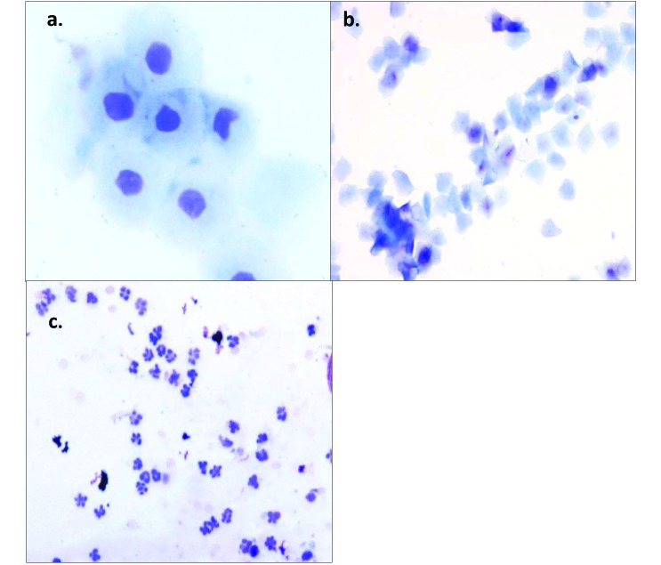
(A) The proestrus phase is characterized by nucleated epithelial cells, which are often found clustered together or in sheets. Magnification, 400×. (B) The estrus phase of the rat is indicated by anucleated cornified cells, which often have angular edges. Magnification, 100×. (C) Diestrus is characterized by an abundance of leukocytes, which often are mixed with cellular debris. Magnification, 200×. Wright–Giemsa stain.

