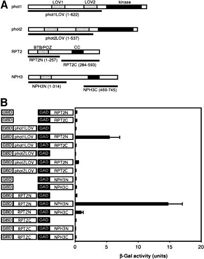
Figure 4.
Interaction between Phototropins and RPT2 or between RPT2 and NPH3.
(A) Schematic of phot1, phot2, RPT2, and NPH3 structures. The protein kinase and LOV domains of phot1 and phot2 proteins are shown as solid and dotted blocks, respectively. The BTB/POZ and coiled-coil (CC) domains of RPT2 and NPH3 are shown as dotted and solid blocks, respectively. Amino acid residues used for each underlined construct are shown in parentheses.
(B) Yeast two-hybrid assay in phot1–RPT2 or RPT2–NPH3 interactions. Solution assays of β-galactosidase (β-Gal) activity were performed for the combinations shown at left. One unit of β-Gal activity was defined as the amount of enzyme that converted 1 μmol of o-nitrophenyl-β-d-galactopyranoside to o-nitrophenol and d-galactose in 1 min at 30°C. Each bar represents an average of 3 to 10 measurements ±sd.

