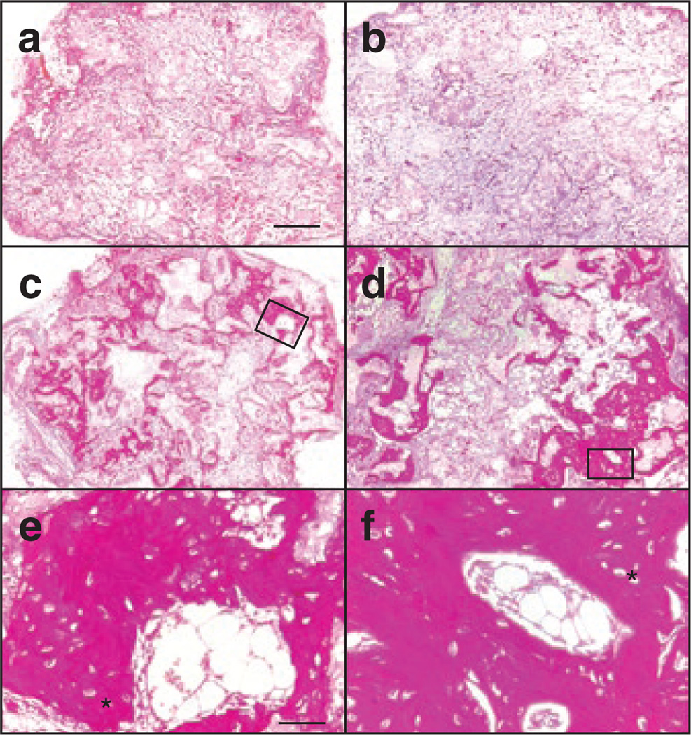
Figure 3. Gene-targeted OIMSCs form bone in immunodeficient mice.
Histological sections of hydroxylapatite/tricalcium phosphate matrices (a) lacking mesenchymal stem cells (MSCs); (b) seeded with normal human fibroblasts; (c, e) OIMSCs targeted at exon 4 of the mutant COL1A2 allele; and (d, f) OIMSCs targeted at exon 2 of the mutant COL1A2 allele, 8 weeks after implantation under the skin of non-obese diabetic/severe combined immunodeficiency mice. Implants were cut into 5 µm sections, stained with hematoxylin and Van Giesonis picric acid fuchsin, and photographed. Regions of dark staining with osteocytes indicate bone. Higher power views are outlined in c and d and shown in e and f, respectively. Lacuna containing osteocytes can be seen, and are indicated by (*). Scale bars = 0.4 mm (a, b, c, d), and 50 µm (e, f).

