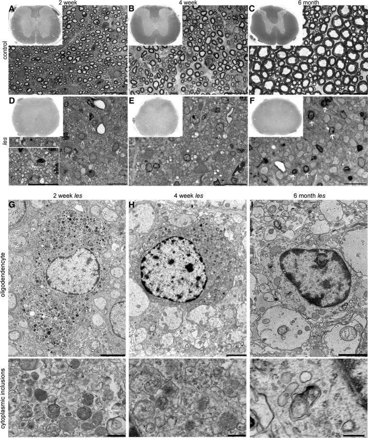
Figure 1.
Representative images of toluidine blue-stained 1 μm sections of control (A–C) and les (D–F) spinal cord. At 2 weeks, les axons develop thin myelin (D, inset). However, most of this myelin is lost by 4 weeks (E), and at later ages myelin is rare in the spinal cord of les rats (F). During myelin development and loss, les oligodendrocytes develop accumulations within their cytoplasm (D,E, arrowhead). At older time points, these cells continue to appear very abnormal (F, arrow). Whole spinal cord images were taken with a 4× objective. The magnified inset at 2 weeks was taken with a 60× objective. Scale bars, 1 mm. Electron micrographs of les oligodendrocytes through myelin development and loss (G–I). At 2 and 4 weeks, les oligodendrocytes are filled with abnormal inclusions, including vesicles, lysosomes, and membrane-bound organelles (G,H, enlarged image). At 6 months, les oligodendrocytes have fewer abnormal inclusions, lack normal Golgi and ER, and have a watery appearance to their cytoplasm (I, enlarged image). Images were taken at 2500× and 5000×. Scale bars: 2500×, 2 μm; 5000×, 500 nm.

