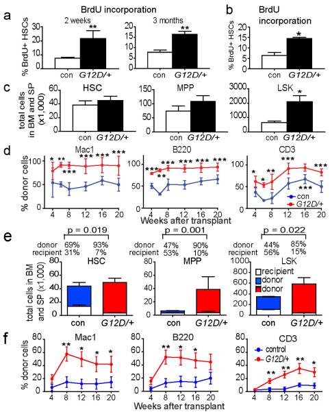
Figure 1. NrasG12D/+ increased HSC proliferation and competitiveness.
a) A 24-hour pulse of BrdU was administered to Mx1-cre; NrasG12D/+ (G12D/+) and littermate control (con) mice at 2 weeks and 3 months after pIpC treatment (n=3 mice/treatment). b) BrdU incorporation by CD150+CD48−LSK HSCs from Vav1-Cre; NrasG12D/+ mice (G12D/+) or littermate controls (con) at 6-10 weeks of age (n=3). c) The total number of CD150+CD48−LSK HSCs, CD150−CD48−LSK MPPs, and LSK cells in the bone marrow and spleens of Mx1-cre; NrasG12D/+ (G12D/+) and littermate control (con) mice at 2 weeks after pIpC treatment (n=5 mice/treatment). d) 5×10⁵ donor bone marrow cells from Mx1-cre; NrasG12D/+ (G12D/+) or littermate control (con) mice at 2 weeks after pIpC treatment (n=3 donors/genotype) were transplanted into irradiated recipient mice (n=15 recipients/genotype) along with 5×10⁵ recipient bone marrow cells. Donor cell reconstitution in the myeloid (Mac-1+ cells), B (B220+), and T (CD3+) cell lineages for 4 to 20 weeks after transplantation. e) Recipients of Mx1-cre; NrasG12D/+ (G12D/+) bone marrow cells (n=5) had significantly (p<0.05) higher proportions of donor-derived HSCs, MPPs and LSK cells compared to recipients of control bone marrow cells (con). f) 10 donor HSCs from Mx1-cre; NrasG12D/+ (G12D/+) or littermate control (con) mice at 2 weeks after pIpC treatment (n=3 donors/genotype) were transplanted into irradiated recipient mice (n=14 recipients/genotype) along with 3×10⁵ recipient bone marrow cells. Data represent mean±s.d.. Two-tailed student's t-tests were used to assess statistical significance. *P<0.05, **P<0.01, ***P<0.001.

