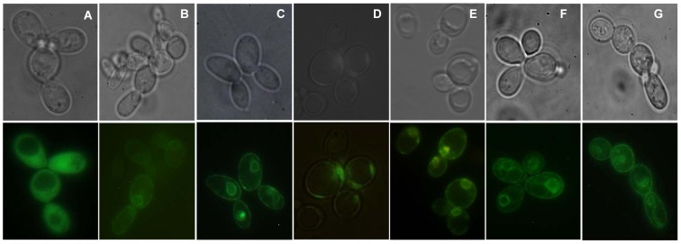
Figure 2. Localization of GFP-tagged aquaporins from V. Vinifera expressed in S. cerevisiae strains.

Cytosolic GFP localization in (A) control cells (transformed with empty plasmid pUG35) and in the membrane of cells expressing (B) VvTnPIP1;4, (C) VvTnPIP2;1, (D) VvTnPIP2;3, (E) VvTnTIP1;1, (F) VvTnTIP2;2, (G) VvTnTIP4;1. Images were taken under phase contrast (upper panel) and fluorescence (lower panel) microscopy.
