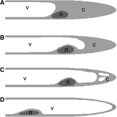
Figure 1.
Cartoon of Root Hair Cytoarchitecture.
Simple representations of the cytoarchitecture of developmental stages of root hairs.
(A) Growing root hair. The subapex of the root hair is filled with cytoplasm (c), and the nucleus (n) is at the base of this area. The shank of the root hair is filled with the central vacuole (v) and cortical cytoplasm.
(B) Early growth-terminating root hair. The first sign of growth termination is that the central vacuole overtakes the nucleus and, therefore, that the subapical region with dense cytoplasm is getting shorter.
(C) Late growth-terminating root hair. The central vacuole expands more and more into the subapex and smaller vacuoles, or extensions of the central vacuole appear into the remaining cytoplasm.
(D) Full-grown root hair. The nucleus has lost its fixed position in the root hair. The vacuole completely fills the root hair and is surrounded by a thin layer of cytoplasm.

