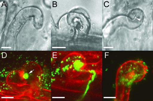
Figure 10.
M. truncatula Wild-Type and dmi2-1 Root Hairs Curl around S. meliloti 2011-GFP.
(A) Reoriented wild-type root hair in the presence of S. meliloti.
(B) Bright-field image of a wild-type root hair curl with an infection thread. Note the complex multifaceted three-dimensional structure of the curl.
(C) Bright-field image of a curled dmi2-1 root hair. Arrowhead points to the furrow at 180° curling.
(D) Projection of 30 images from a confocal laser scanning microscope Z-stack of a wild-type curled root hair, entrapping a GFP-expressing bacterial colony (arrow). The cell wall (red) was counterstained with 0.1% propidium iodine. Note the multifaceted three-dimensional structure of the root hair curl.
(E) Projection of 35 images of a Z-stack from a wild-type curled root hair with a bacterial colony in a closed pocket.
(F) Projection of 20 images of a Z-stack from a dmi2-1 root hair, curling in the presence of bacteria but unable to entrap them. Note the bacteria on the outside of the root hair. Monitored were four roots per wild type and dmi2-1. Bars = 15 μm.

