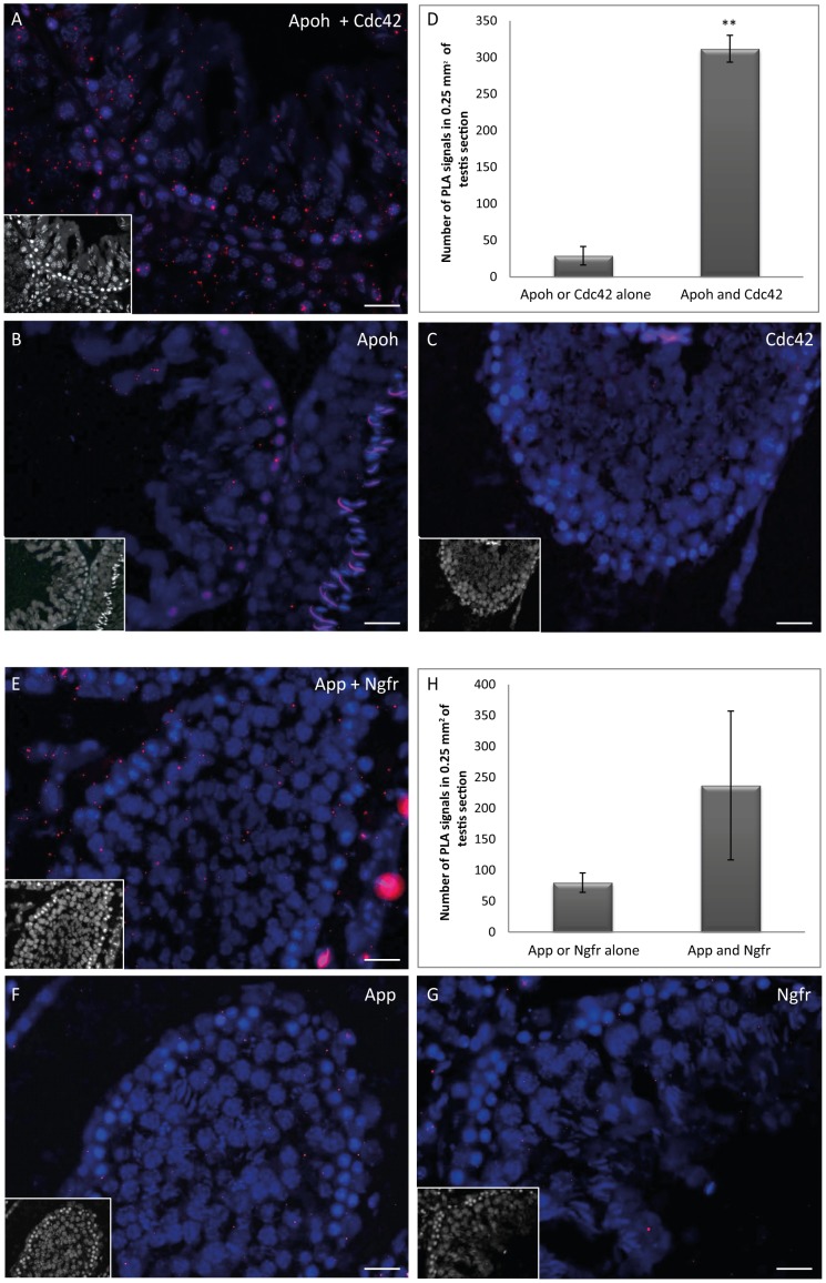
Figure 4. In situ detection of APOH/CDC42 and APP/NGFR interactions in the rat testis by Duolink PLA.
(A, E) Abundant PLA signals (red dots) were detected in the seminiferous tubules of rat testis when specific primary antibodies were used, reflecting the close proximity of APOH/CDC42 or APP/NGFR close proximity. (B, C, F, G) Negative controls, with only one primary antibody for the targeted protein-protein interaction. (D, H) Quantification of PLA signals in testis sections for APOH/CDC42 (** Student's t-test p = 0.0016) or for APP/NGFR (Student's t-test p = 0.16). Scale bars = 50 µm. A nonspecific background nuclear signal was observed for a few tubule sections (see B).

