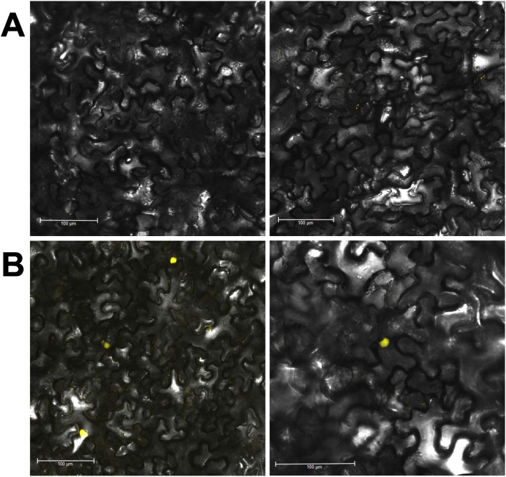
Figure 7. Epidermal cells of N. benthamiana expressing translational fusion of StMKK6 with YFP under native promoter.
Leaves were agroinfiltrated when the virus has spread uniformly through the inoculated leaves (8 days after inoculation) and observed after 72 h in two independent experiments. Examples from two plants (left and right panels) are shown. Control of transformation (fluorescent marker without StMKK6 fusion) is in the Figure S1A. A. Localisation of StMKK6 in mock-inoculated leaves. No fluorescence was observed. B. Localisation of StMKK6 in PVY-inoculated leaves, where the protein accumulates predominantly in nucleus. Additional images of StMKK6 localisation under native promoter are in Figures S1D and S1E.

