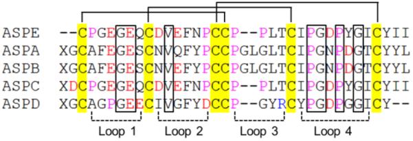
Figure 2.

Sequence alignment of ASPE with asteropsins A–D (ASPA, ASPB, ASPC, and ASPD). The disulfide bonds are connected by solid lines. The loops indicate residues that compose polypeptide backbones between two Cys residues. Residues: red = acidic, yellow = Cys, purple = Pro. X = pyroglutamic acid. Conserved structure-maintaining residues are marked with boxes. Sequence alignment was performed using ClustalX 2.0 (BMBL-EBI).
