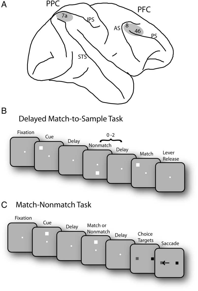
Figure 1.
Brain areas and tasks. (A) Schematic diagram of the monkey brain. The areas of recordings are highlighted. AS, arcuate sulcus; IPS, intraparietal sulcus; PS, principal sulcus; STS, superior temporal sulcus. (B) Successive frames illustrate the sequence of stimulus presentations in the Delayed Match-to-Sample task. Following the cue presentation, a match or nonmatch stimulus appeared. The monkeys were required to remember the location of cue stimulus and release a lever when a subsequent stimulus appeared at the cued location. (C) Schematic illustration of the Match/Nonmatch task. Two choice targets were presented at the end of a trial, and the monkey was required to saccade to a green target (colored in gray in the figure); in case the 2 stimuli were matching and to a blue target (colored in black) otherwise.

