Figure 12.
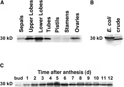
GPPS.SSU Protein Levels in Different Floral Tissues and in Petals during Flower Development.
(A) Expression of GPPS.SSU protein in sepals, upper and lower petal lobes, tubes, pistils, stamens, and ovaries of 4-d-old A. majus flowers. Representative protein gel blot shows the 30-kD protein recognized by anti-GPPS.SSU antibodes. Proteins were extracted from different floral tissues, and 20 μg of protein was loaded in each lane. The blot shown represents a typical result of two independent experiments.
(B) Immunodetection of GPPS.SSU in the recombinant GPPS and in crude petal extract.
(C) Expression of the GPPS.SSU protein in upper and lower petal lobes at different stages of development. Proteins were extracted from upper and lower petal lobes at different stages of development, and 20 μg of protein was loaded in each lane. The blot shown represents a typical result of six independent experiments.

