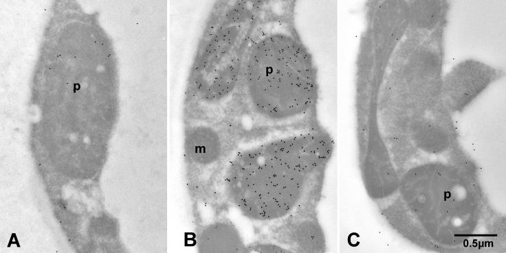
Figure 8.
Intracellular Localization of GPPS.SSU in A. majus.
(A) Transmission electron microscopy (TEM) image of conical cells of 1-d-old A. majus lower petal lobe labeled with anti-GPPS.SSU antibodies and gold-conjugated goat anti-rabbit antibodies. p, plastid.
(B) TEM image of conical cells of 7-d-old A. majus lower petal lobe labeled with anti-GPPS.SSU antibodies and gold-conjugated goat anti-rabbit antibodies. m, mitochondria; p, plastid.
(C) TEM image of conical cells of 7-d-old A. majus lower petal lobe treated with preimmune serum and gold-conjugated goat anti-rabbit antibodies. p, plastid.

