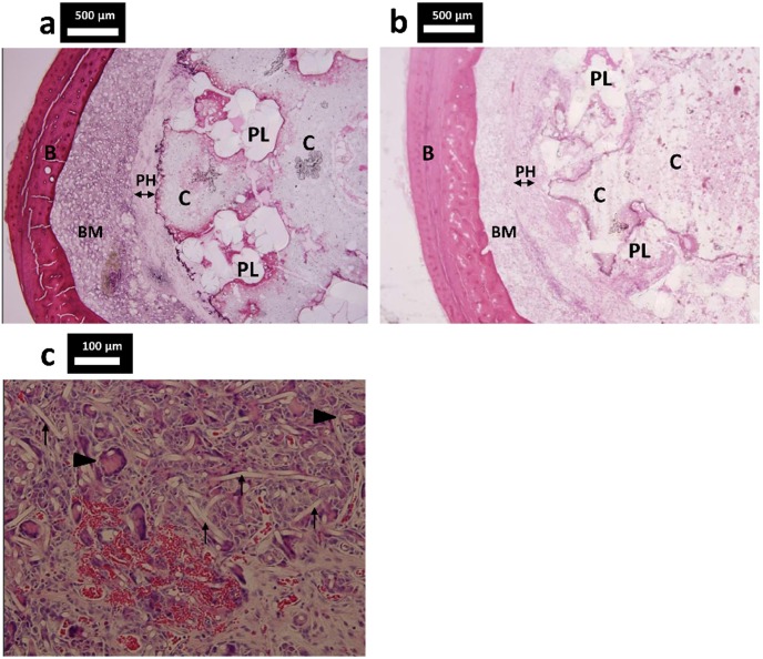
Figure 8. Histologic cross sections of specimens from the IM-BM group.
Histologic cross sections of specimens from the IM-BM group (PLLA + CPC + PHA) stained with hematoxylin and eosin. (a) A PHA fiber mat layer surrounded the CPC, and the PLLA tube was not degraded at week 20. (b) At week 52, the PLLA tube seemed to have degraded gradually. (c) Multinucleated giant cell (arrowhead) and neovascularization were observed in the PHA fiber mat layer at week 52. PHA fiber (arrow). PL: PLLA woven tube; PH: PHA fiber mat C: CPC; B: bone cortex; BM: bone marrow.

