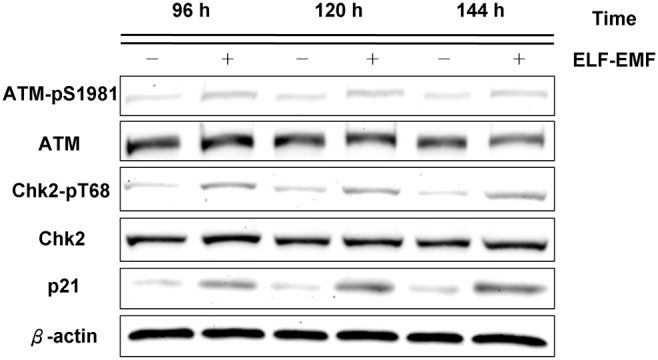
Figure 3. Immunoblotting of phospho-ATM (Ser1981), phospho-Chk2 (Thr68), p21 after ELF-EMF exposure in HaCaT cells.

The expression levels of phospho-ATM (Ser1981), phospho-Chk2 (Thr68), and p21 in the exposed cells were higher than those in the sham-exposed cells at the indicated times. β-actin was used as a loading control. All proteins were determined in whole cell lysates from the sham and exposed HaCaT cells after the indicated exposure times.
