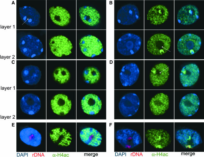
Figure 4.
rDNA Repeats Are Hyperacetylated in Nuclei of HDA6 Mutants.
(A) to (D) Distribution of histone H4 acetylation revealed by DAPI staining of DNA (blue, left panel) and immunodetection with an antibody specific for tetra-acetylated H4 (green, middle panel) in nuclei of control lines DR5 (A) and Ler (C) and in axe1-5 (B) and sil1 (D) mutant nuclei. Right panels show merged images. For each nucleus, two layers were selected from deconvoluted image stacks, arrows mark the nucleolus.
(E) and (F) FISH using rDNA repeats (red, left panel) after immunostaining with α-H4ac antibodies (green, middle panels) shows that the rDNA loci indeed are devoid of H4ac staining in the wild type (E) but become highly enriched with H4ac in mutant nuclei (F).

