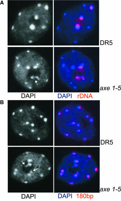
Figure 7.
rDNA Loci, but Not Chromocenters in General, Are Decondensed in HDA6 Mutant Nuclei.
Interphase nuclear spreads of control lines DR5 and Ler and mutants axe1-5 and sil1 stained with DAPI (black and white in left panel, blue in merged images in the right panel) and FISH with biotin-labeled probes for rDNA repeats (A) and centromeric (180 bp) repeats (B). Arrows in the black and white images point to decondensed rDNA repeats in mutant nuclei in (A) and (B).

