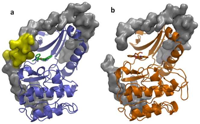
Figure 4. Disordering of the PRK1 C-tail in the lestaurtinib crystal structure.

(a) PRK1:Ro-31-8220 is shown with the protein in blue, with the inhibitor in green. The C-tail is shown as a surface in gray; the region disordered in the lestaurtinib structure is shown in yellow on the Ro-31-8220 structure. (b) PRK1:Lestaurtinib is shown colored orange with the C-tail shown as a surface in grey, with a gap visible due to disorder of the C-tail proximal to the inhibitor.
