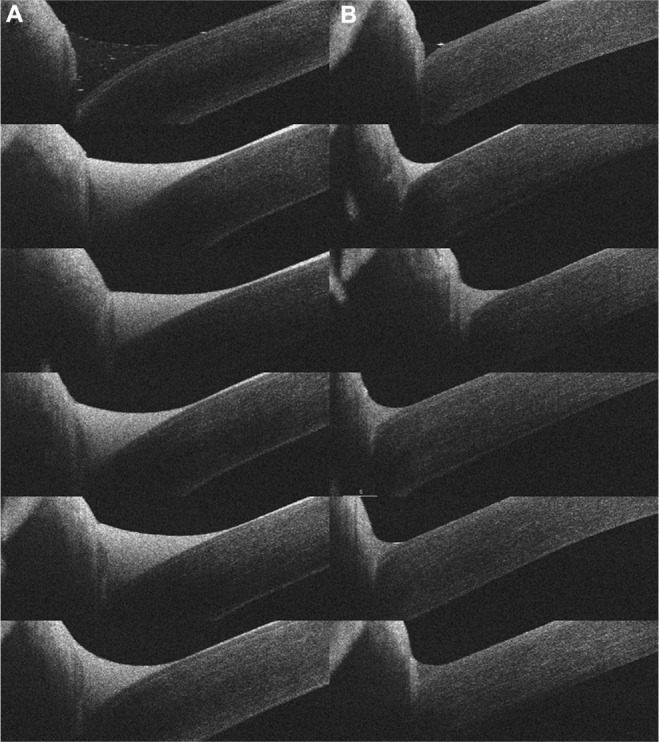
Figure 1.

Optical coherence tomography images of the tear meniscus.
Notes: Serial images of the tear meniscus before (A) and after (B) an operation on a 70-year-old woman. The tear meniscus was measured before (Top) and at 1, 3, 5, 7, and 10 minutes (Bottom) after rebamipide instillation.
