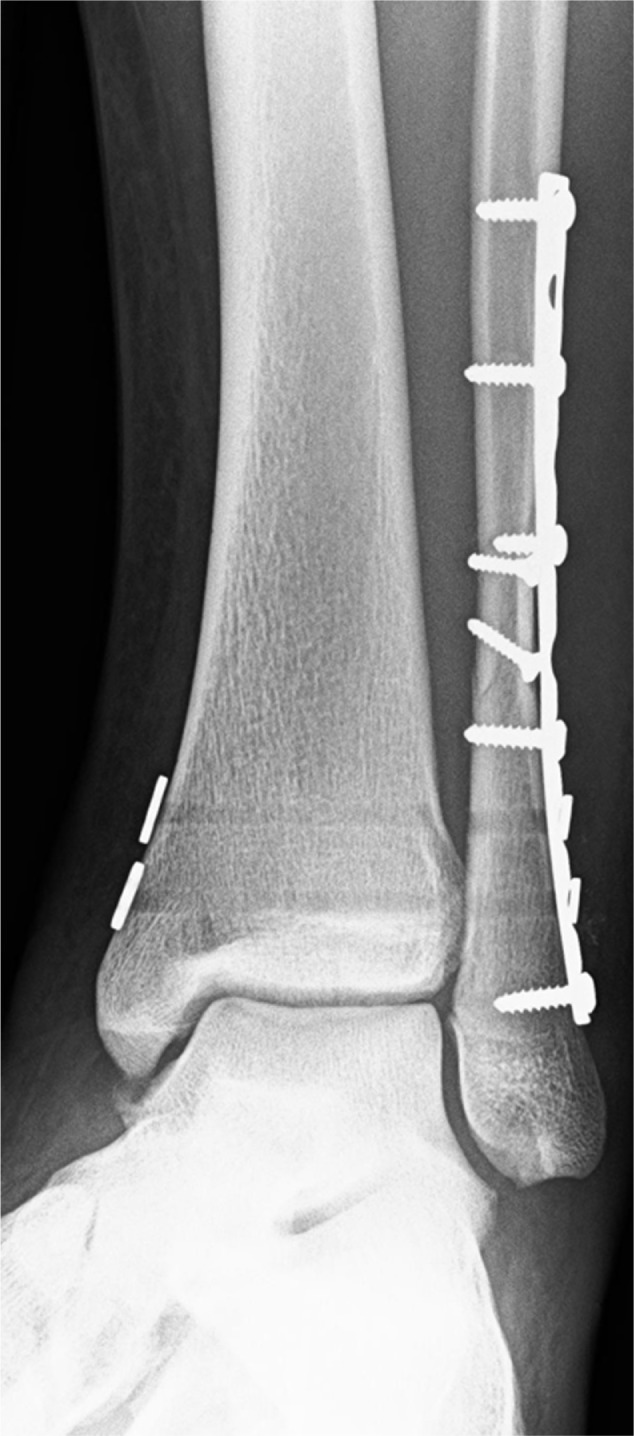
Figure 2.

Postoperative left anterior-posterior ankle radiograph after open reduction and internal fixation of Weber C fibular shaft fracture with lag screws, one third tubular plate and two suture button fixation of the syndesmosis demonstrating anatomic alignment of the syndesmosis, medial clear space and fibular shaft. The deltoid ligament was also repaired in this athlete.
