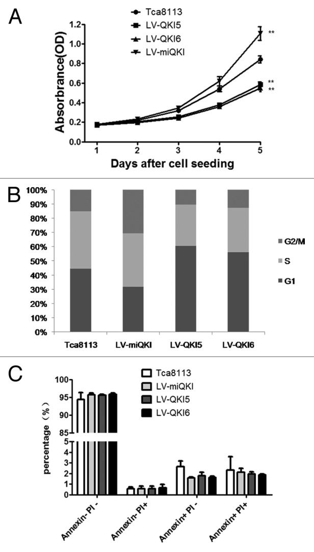
Figure 3. QKI inhibits the expansion of OSCC cells and has no marked effect on cell apoptosis. (A) Compared with the control cells, overexpression of QKI5 or QKI6 significantly inhibited the growth of Tca8113 cells, and knockdown of QKI promoted the growth of Tca8113 cells. LV-QKI5, LV-QKI6, LV-miQKI and Tca8113 cells were cultured for indicated time and cell number were analyzed by MTT (n = 5, **P < 0.01). (B) Cell cycle distribution of cells with QKI overexpression and knockdown. Results from flow cytometric analysis showed that compared with the control cells, the proportion of cells in S and G2/M phase was increased in LV-miQKI cells, and the proportion of cells in G1 phase was increased in LV-QKI5 and LV-QKI6 cells. Data presented here is a representative of 3 different experiments. (C) Flow cytometric analysis showed no marked effect on cell apoptosis. Data from 3 independent experiments are presented as mean ± SD. PI, propidium iodide.
