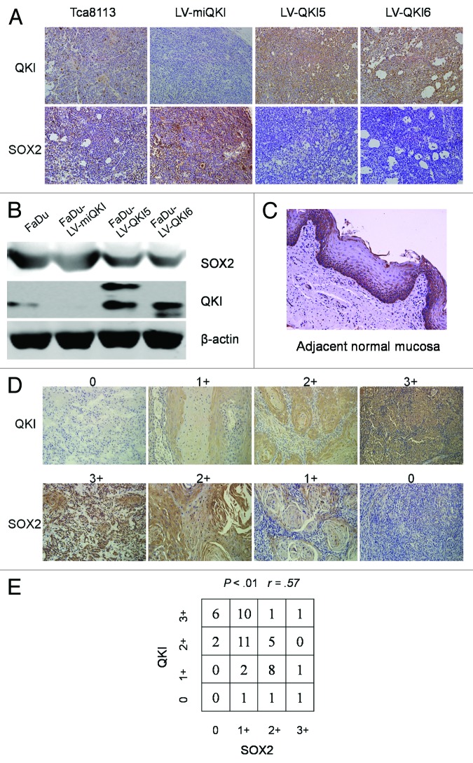
Figure 5. QKI causes decreased SOX2 expression at mRNA and protein level. (A) Immunostaining of QKI and SOX2 was performed on subcutaneous tumors inoculated with 1 × 10⁶ cells after 6 wk (original magnification 200×). Tumors derived from LV-QKI5 or LV-QKI6 cells displayed lower SOX2 immunostaining than the control, and tumors derived from LV-miQKI cells displayed higher SOX2 immunostaining than the control. (B) Expression of QKI and SOX2 in FaDu cells, and FaDu cells infected with LV-QKI5, LV-QKI6, or LV-miQKI was detected by western blot. (C) Representative data of the expression of SOX2 in adjacent normal mucosa assayed by immunohistochemistry (original magnification 200×). SOX2 was highly expressed in the stratum basale which contained stem cells. (D) Representative QKI and SOX2 expression in oral cancer tissue microarray sections of 50 patients (original magnification 200×). The immunostaining levels of QKI and SOX2 were scored from 0 to 3. (E) Correlation of expression levels of QKI and SOX2 is shown (r = 0.5714, P < 0.01).

An official website of the United States government
Here's how you know
Official websites use .gov
A
.gov website belongs to an official
government organization in the United States.
Secure .gov websites use HTTPS
A lock (
) or https:// means you've safely
connected to the .gov website. Share sensitive
information only on official, secure websites.
