
Fig. 3.
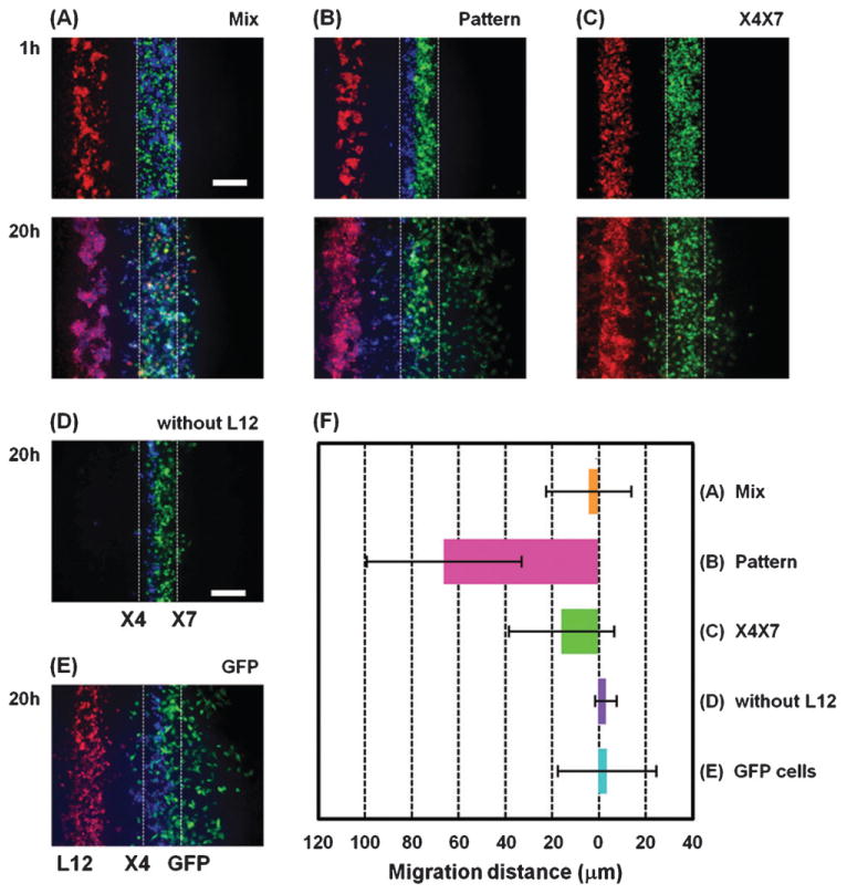
Effect of closely positioning CXCR4 and CXCR7 expressing cells together. (A, B) X4 cells (blue) and X7 cells (green) were co-patterned over the middle channel of the lower layer by either randomly mixing together (A) or by patterning side-by-side (B). In (C) MDA-MB-231 cells stably expressing both CXCR4 and CXCR7 (green) were patterned over the middle channel of the lower layer. For comparison, X4 cells (blue) were patterned side-by-side to X7 cells (green) without L12 cells (red) in (D) and X4 cells (blue) and GFP cells that do not express CXCR7 (green) were co-patterned over the middle channel of the lower layer in a side-by-side manner in (E). Fluorescent images were taken 1 h and 20 h after seeding the cells. (F) Quantification of X4 cell migration under each condition (n = 5–10 independent experiments for each condition). The X-axis shows distance from the center of the initial position of X4 cells. Scale bars: 200 μm.