
Figure 1.
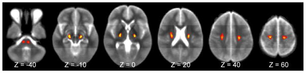
Density-weighted CST atlas in MNI152 space. Several axial (z-level) images from the level of the pons to precentral gyrus are shown. Voxels are color-coded for the relative density (scale of 0–100) of CST streamlines based on tractography of DTI data from healthy subjects.