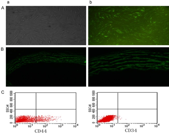
Figure 3.

Identification of bone marrow mesenchymal stem cells and differentiation to vessels. Cultured primary bone marrow mesenchymal stem cells attached to the walls and became spindle-shaped, some assuming polygonal pattern, and grew in clusters. Single cells were occasionally observed. At day 5, obvious colonies formed, and at day 10 almost all cells attached to the walls, being flat and narrow and assuming vortex pattern (Aa). Three days after infection with slow virus, green fluorescent signals were observed under fluorescent microscope (Ab). Flow cytometry showed that 0.19% of the cultured cells were CD34-negative and 25.25% of the cells were CD44-positive. The findings confirmed that the cultured cells of 3-4 passages were bone marrow mesenchymal stem cells (C). After transplantation of the stem cells, in MSC group, MSCs were found to have migrated to and settled in cardiac tissues, eventually differentiating into blood vessels (B: a: 200× b: 400×).
