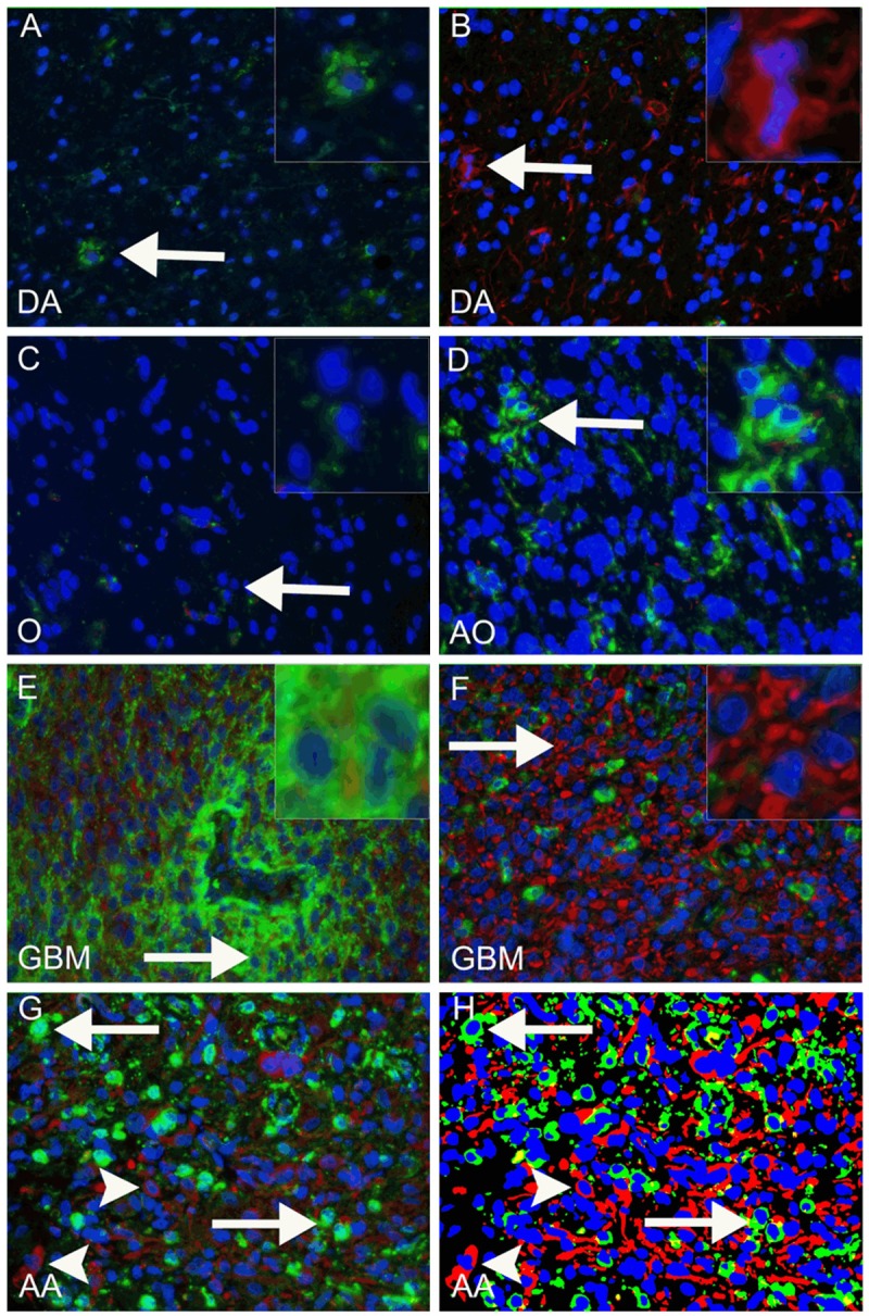
Figure 1.

Fluorescence-based staining identifying CD133 and nestin. Examples of CD133-positive areas in diffuse astrocytomas (DA), oligodendrogliomas (O), anaplastic oligodendrogliomas (AO), glioblastomas (GBM) and anaplasticastrocytomas (AA) are shown in (A, C-F) (green, arrows indicate site of inserts). In E a GBM with a CD133 positive peri-vascular niche is shown. Examples of nestin-positive areas are shown in (B and F) (red, arrows indicate site of inserts). When the original fluorescence image (G) was processed and the algorithm applied (H), CD133 positive staining was shown in green, nestin positive staining in red and nuclei in blue. Arrows in G and H indicate CD133 positive cells, arrowheads indicate nestin positive cells.
