
Figure 5.
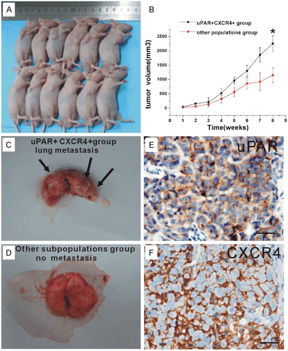
The xenograft and lung metastasis forming of uPAR+CXCR4+ and other subpopulation cells in vivo. A: Subcutaneous implantation of 5 × 10⁵ uPAR+CXCR4+ cells or other subpopulation cells in BALB/c nude mice led to tumor formation (6/6, 5/6, respectly). B: BALB/c nude mice shows that after 8 weeks injected, growth curve of xenograft tumors volume of uPAR+CXCR4+ group exhibit significant larger than other subpopulation group (2227.6 ± 211.35, 1201.0 ± 170.36 mm3, respectively, P < 0.01). C: After 8 weeks, mice were sacrificed, lung metastasis foci were founded in uPAR+CXCR4+ cells group (arrow indicated). D: Other subpopulation cells group showed no trace of metastasis. E: Immunohistochemistry showed uPAR positive expressed in lung metastasis tumor tissue. F: Immunohistochemistry showed CXCR4 positive expressed in lung metastasis tumor tissue (× 400, Scale bar = 50 μm).