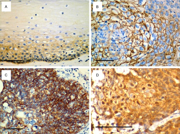
Figure 1.
Representative immunohistochemical analysis of β-catenin expression. (A) Normal cervical epithelial cells displayed membranous and cytoplasmic expression of β-catenin in the basal and suprabasal layers. (B) Membranous, (C) membranous and cytoplasmic and (D) nuclear patterns of β-catenin expression were observed in cervical squamous cell cancer (CSCC). Scale bar, 100 μm.

