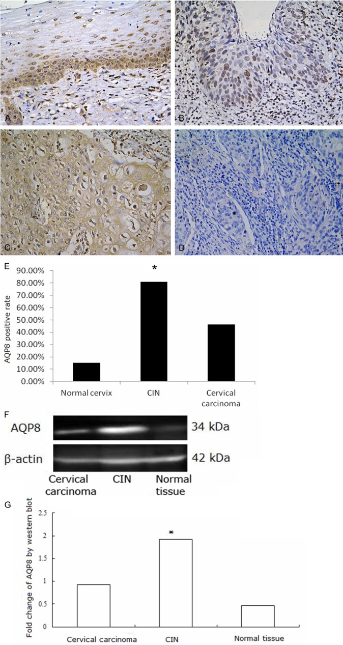
Figure 2.

AQP8 expression in cervical tissues. A: AQP8 expressed in the membrane and cytoplasm of basal and proliferative prickle cells in normal cervical tissue; B: AQP8 expressed in the nuclei in CIN; C: AQP8 expressed in the cytoplasm and membrane in cervical cancer; D: Negative control of AQP8 expression in cervical cancer; E: Fraction of AQP8 positive cells in each group. A-E: AQP8 expression detected by Immunohistochemistry. Magnification × 200. *P < 0.05. F, G: AQP8 expression detected by Western blot was higher in CIN than in cervical carcinoma or normal tissue samples *P < 0.05.
