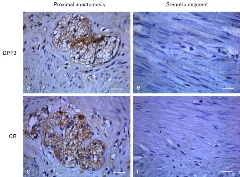
Figure 4.

Immunohistochemical staining analyzed the protein expressions of DPF3 and CR in different segments of HSCR (400 ×). Intensive stainings of DPF3 (A) and CR (C) were displayed in ganglion cells and myenteric plexuses of proximal anastomosis. In the stenotic segment of HSCR, positive stainings of DPF3 (B) and CR (D) were not observed between the circular and the longitudinal muscle. Scale bar = 100 μm.
