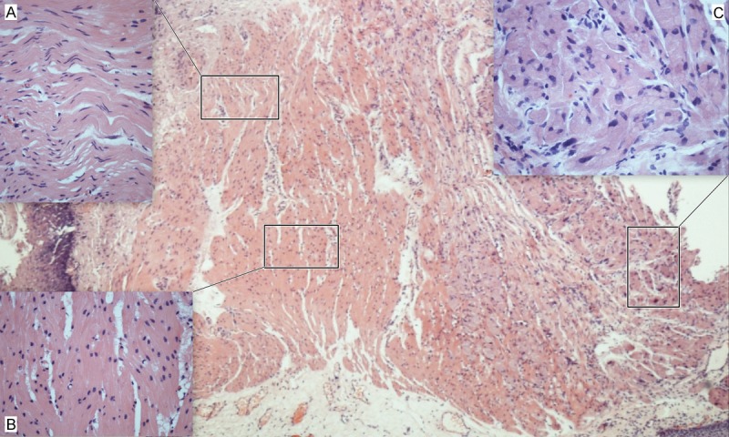
Figure 3.

A developmental morphology of a GCT of esophagus. It included areas of (A) relatively normal Schwann cells, (B) transitional cells and (C) typical cells of GCTs. The transitional cells generally kept the arrangement pattern of normal Schwann cells, however, the nuclei turned oval and round, and the cytoplasm turned to be granular.
