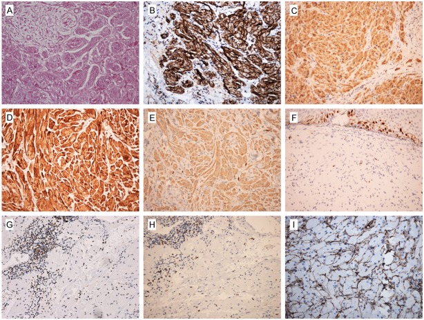
Figure 4.
Immunohistochemistry (IHC) and histochemistry staining results of GCTs of esophagus. (A) PAS staining was positive and diastase resistant. Tumor cells were moderate to strong positive staining of (B) Nestin, (C) NSE and (D) S100 protein. (E) CD68 was moderate positive. (F) Ki67 had a low positivity. The scattered lymphocytes in tumor stroma or the hyperplastic lymphoid tissues around the tumor were showed positivity of (G) CD45RO and (H) CD8. The stroma around the tumor cells were positive for (I) CD34.

