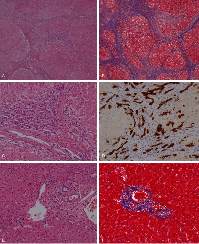
Figure 1.

Microscopic findings well-defined regenerative nodules surrounded by thick fibrous septa with collagen deposition are seen in H & E (A) and Masson’s trichrome (B) stain. Cholangiocytes of bile ductule proliferate in septal area (C) demonstrated with CK7 immunohistochemically (D). In control cases, well-formed portal triads which consist of vein (portal vein), artery (hepatic artery) and biliary tract are seen (E). Collagen deposition is observed only in portal area and its close periphery (F). (A, C, E) Hematoxylin and eosin; (B, F) Masson’s trichrome; (A and B) x 4; (C to F) x 200.
