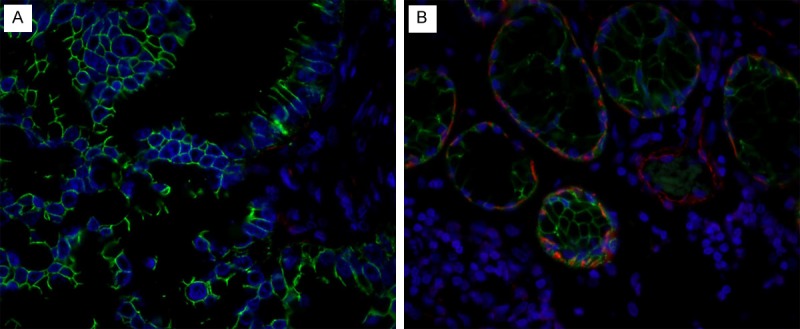
Figure 2.

Double immunofluorescence staining of E-cadherin (detected in green) and α-SMA (detected in red) in the tumor tissue at the center (A) and the border (B) of the primary adenocarcinoma in lung. (A) E-cadherin positive cells are detected, but α-SMA is not detected in the center of the tumor; (B) Note that both E-cadherin positive and α-SMA positive cells are detected at infiltrating border of tumor. This is associated with the lymph node metastasis of the tumor cells. The blue staining represents the nuclei (original magnification X 400).
