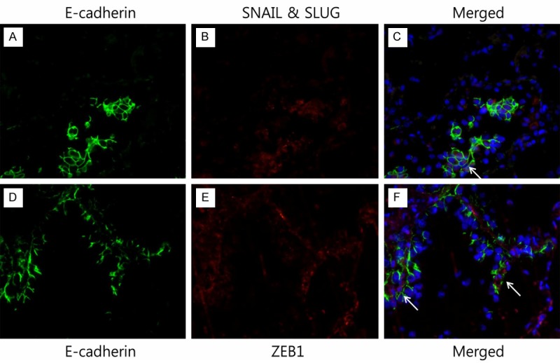
Figure 3.

Merged images of double immunofluorescence multi-staining of the tumor tissue of lung adenocarcinoma. (A, D) anti-E-cadherin antibody, (B) anti-SNAIL antibody, and (C) merged image E-cadherin/SNAIL & SLUG. The coexpression of E-cadherin and SNAIL & SLUG is noted (marked with arrow). (E) Anti-ZEB1 antibody, and (F) merged image E-cadherin/ZEB1. The coexpression of E-cadherin and ZEB1 is noted (marked with arrow). The blue staining represents the nuclei (original magnification X 400).
