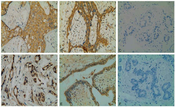
Figure 1.

Immunohistochemical analyses of ALDH1 and TGFβ2 expression in sections of different breast tissues. A: Breast cancer cells showed extensive cytoplasmic staining for ALDH1. B: Cytoplasmic positive staining for ALDH1 in benign fibroadenoma. C: ALDH1-negative staining in non-cancerous normal tissue adjacent to cancer. D: Strong expression of TGFβ2 in the cytoplasm of breast cancer cells. E: Diffuse cytoplasmic staining for TGFβ2 in fibroadenoma tissue. F: TGFβ2-negative staining in paracancerous normal tissue. Representative immunohistochemical examples of staining were shown (original magnification, ×400).
