
Figure 3.
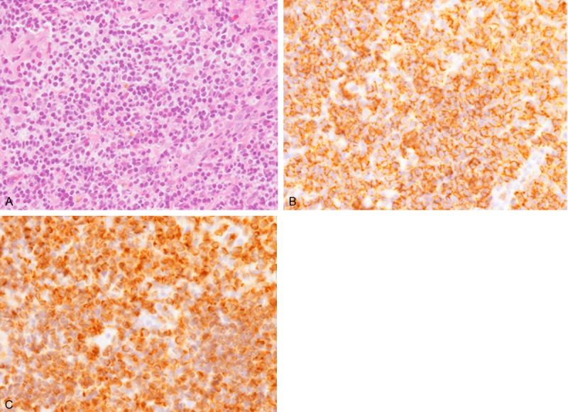
Histopathological and immunohistochemical features of the submucosal tumor of the esophagus. A. Diffuse proliferation of small- to medium-sized lymphocytes with convoluted nuclei and inconspicuous nucleoli. Some of them have clear cytoplasm. HE, x 400. B. CD20 is diffusely expressed in these lymphocytes x 400. C. Bcl-2 is also diffusely expressed x 400.