
Figure 2.
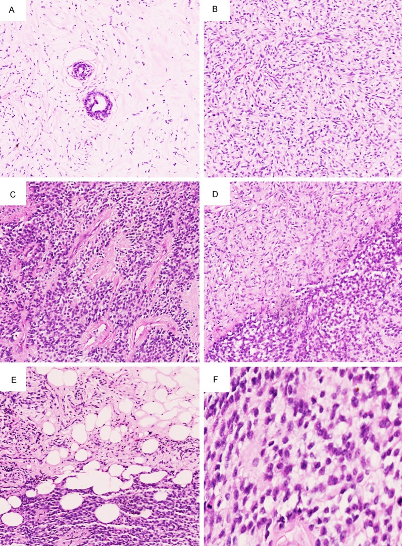
Microscopic features of breast malignant SFT. A. In hypocellular area, a small mount of spindle-shaped tumor cells scattered in collagen fibers, extremely rare mammary gland-like structure could be seen, HE 100 ×. B. In hypercellular area, Ovoid-spindle shaped tumor cells surrounded branching and staghorn vasculature, HE 100 ×. C. Round shaped tumor cells showed hemangiopericytoma-like growth pattern, HE 100 ×. D. There is a relative clear boundary between hypocellular and hypercellular areas, HE 100 ×. E. Focally, infiltrative margins could be identified, HE 100 ×. F. Tumor cells showed mild to moderated cytological atypia and active mitoses, HE 400 ×.