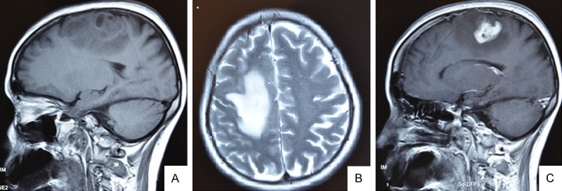
Figure 1.

MR Images of this case displaying a bulging lesion with marked peripheral edema localized on the right frontal region. MRI showing a T1-isointensity (A), T2-heterointensity (B) and the central part of lesion was observed after contrast medium injection (C). (A, C, sagittal view; B and A, axial view).
