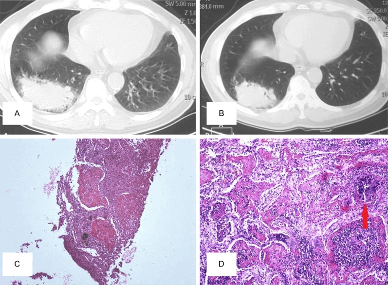
Figure 2.

A: In case 2, CT-scan discovered an occupying mass under the pleura in the lower lobe of the right lung with irregular boundaries. B: 3 months after treated with antibiotic and steroids, CT-scan re-examination showed a slight shrink of the previous lesion size. C: Massive intra-alveolus fibrinous deposition with severe acute and chronic inflammatory cells infiltration (original magnification *100) was observed in the biopsy tissue. D: Lobectomy specimen examination discovered a poorly-differentiated adenocarcinoma, intra-alveolus fibrin balls irregularly mixed with cancer cells (original magnification *200, red arrowed pointing at poorly-differentiated adenocarcinoma).
