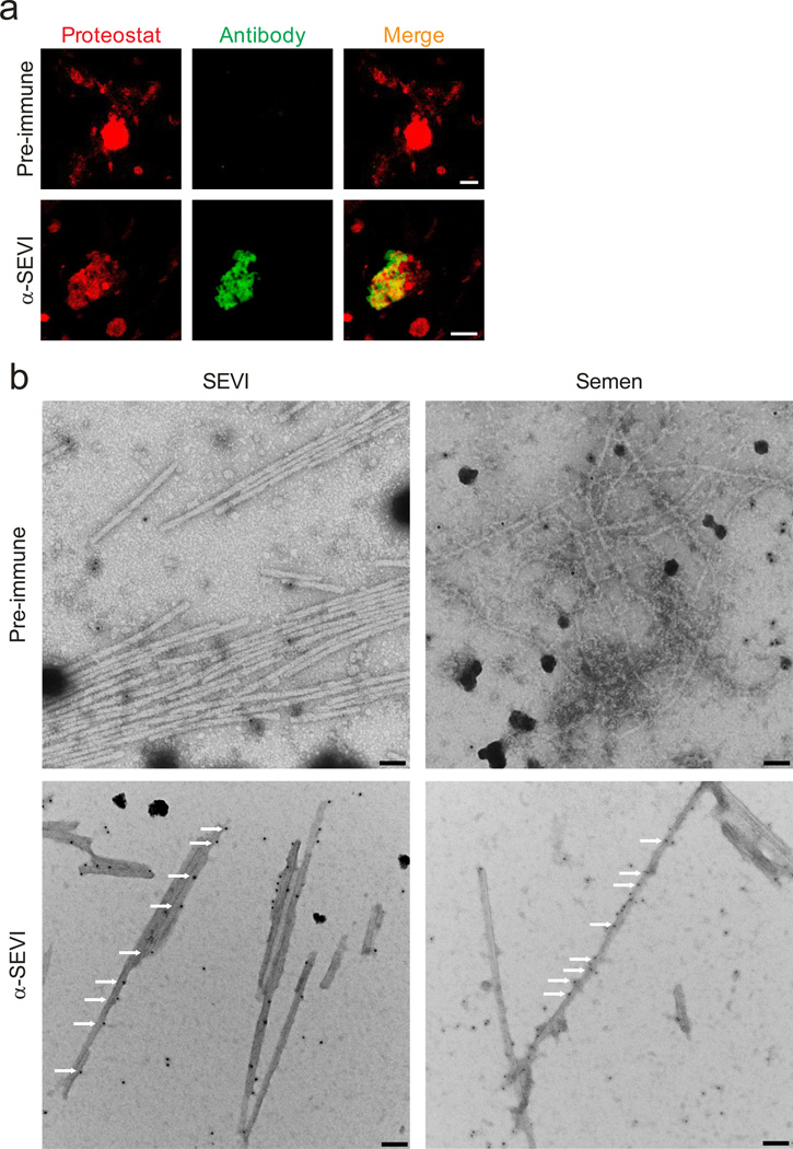
Figure 3. Endogenous amyloids partially consists of SEVI.
(a) Semen was treated with pre-immune (top) or anti-SEVI antiserum (α-SEVI, bottom). The amyloid/antibody complexes were pelleted, washed, and incubated with an Alexa488-coupled secondary antibody (green), and counterstained with Proteostat dye (red). Scale bar = 5 µm. (b) Immunogold-labeling of endogenous SEVI fibrils in semen. Transmission electron micrographs of semen treated with a pre-immune serum or an anti-SEVI antiserum as primary antibodies, and gold conjugated anti-rabbit secondary antibody. Scale bar = 100 nm. White arrows indicate gold particles bound to amyloid fibrils.

