Fig. 2.
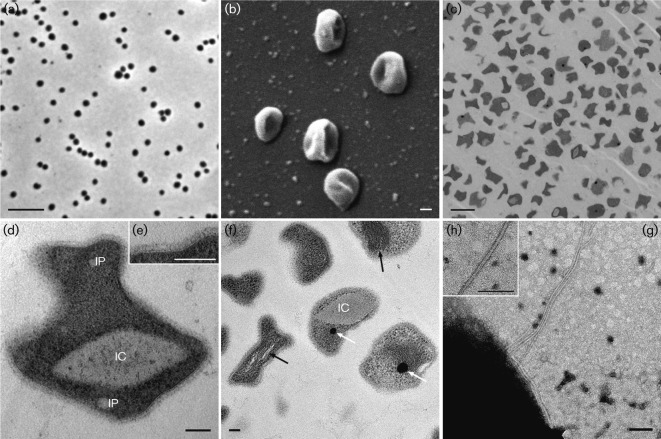
Ultrastructure of cells of strain EN76T. (a) Phase-contrast image; bar, 5 µm. (b) Scanning electron micrograph of several cells depicting the irregular coccoid shape; bar, 100 nm. (c–f) TEM images of ultrathin sections of chemically fixed cells of strain EN76T. (c) Overview displaying the irregular cell shape; bar, 1 µm. (d) Magnified cell showing intracellular features including a clearly discernible area [potential intracellular compartment (IC)] and incorporations (IP). The inset (e) illustrates the cell membrane, pseudo-periplasm and S-layer at higher magnification; bars, 100 nm. (f) Potential intracellular compartment (IC), tubule-like structures (white arrows) and electron-dense particles (black arrows) are highlighted; bar, 100 nm. (g, h) Transmission electron micrographs of a cell with an archaellum; inset (h) shows the magnified archaellum. Bars, 100 nm.

