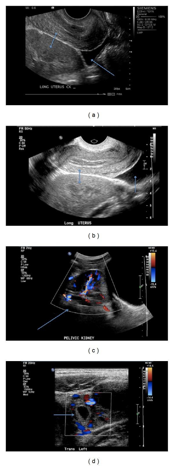
Figure 1.

(a) Longitudinal view: an empty uterus superiorly and pelvic kidney inferiorly (double arrow), free fluid in the cul-de-sac (single arrow). (b) Longitudinal view: an empty uterus superiorly and pelvic kidney inferiorly (double arrow), free fluid in the cul-de-sac (single arrow). (c) Longitudinal view: ectopically located pelvic kidney with color Doppler (single arrow). (d) Transverse view: hypoechoic cystic structure with vascular flow in the left adnexa depicting a tubal pregnancy (single arrow).
