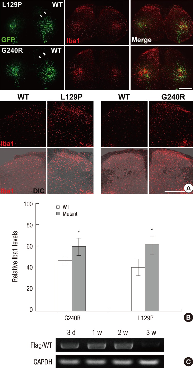
Fig. 2.
Increased number of microglia in the GARS-mutant-expressed dorsal horn. (A) Increase in the number of Iba1-positive microglia in the dorsal horn of the L5-spinal cord 7 days after the transfection of adenovirus vectors into sciatic nerves. GFP expression (green) and immunofluorescence labeling of Iba1 (red) were identified in spinal cord cross-sections. Lower panels are higher magnification images of the spinal dorsal horn. Middle two panels indicates the high magnification images of the upper panels (arrows). Scale bar = 500 µm. (B) Quantification of activated microglia in the dorsal horn at Day 7. The number of Iba1 was calculated with the correction using GFP-neuronal bodies in spinal cord. *P < 0.001 compared with GARS WT (WT, n = 5; L129P, n = 5; G240R, n = 4). (C) mRNA expression of FLAG-tag in the spinal cord following infection with GARS-WT-expressing adenoviruses by RT-PCR.

