
Figure 5.
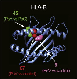
3D Ribbon Models for HLA-B
HLA-B structures are based on Protein Data Bank entry 2bvp36 and were prepared with UCSF Chimera version 1.7.37 Amino acid position 67 and 9 residues associated with overall PsV risk are highlighted as red and pink spheres, respectively. An amino acid position 45 residue associated with subphenotype risk heterogeneity (PsA versus PsC) is highlighted as a green sphere.