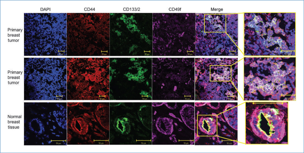
Figure 4.
Localization of CD44, CD49f, and CD133/2 in primary ER-negative breast tissue and normal breast tissue. Confocal microscopy images of representative patient tumors and a normal breast sample illustrating localization of DNA (DAPI; blue), CD44 (red), CD133/2 (green), and CD49f (purple). In the merged images, cells simultaneously positive for all three markers appear white.

