
Figure 3.
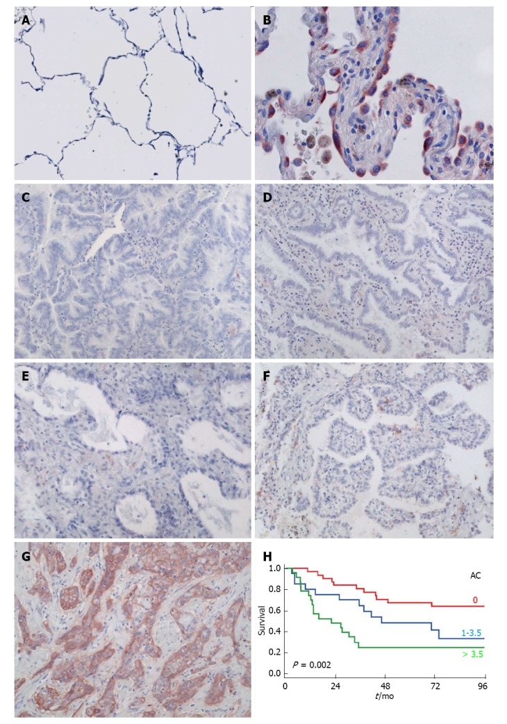
C4.4A in pulmonary adenocarcinoma. Panels A-E: C4.4A expression as detected by immunohistochemistry with a polyclonal antibody in normal alveoli (A; reproduced with permission from Jacobsen et al[34], 2012), atypical adenomatous hyperplasia (B), invasive AC with predominant mucinous lepidic (C), non-mucinous lepidic (D), acinar (E), papillary (F) and solid (G) pattern. Panel H: Kaplan-Meier estimates for the survival of AC patients, which is correlated with C4.4A scores, here stratified by tertiles (red: Lowest level of C4.4A; blue: Intermediate level of C4.4A; green: Highest level of C4.4A). Modified from Jacobsen et al[33], 2013.