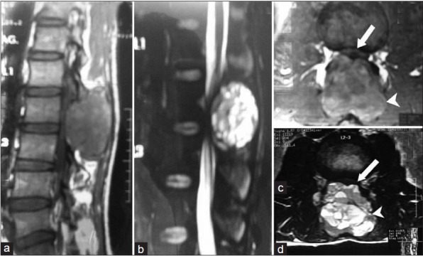
Figure 2.

MRI spine showing well defined expansile mass lesion (arrow head) at L2 vertebra with bilateral laminar destruction and tecal sac compression (arrow). It displayed hypo intense signals on T1; (a and c) Hyper intense signals on T2 weighted images; (b and d) Multiple internal septations and heterogeneous contrast enhancement
