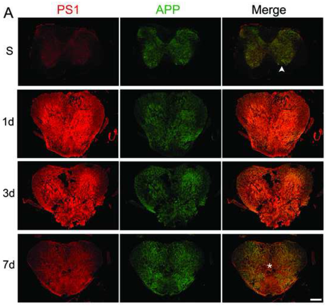
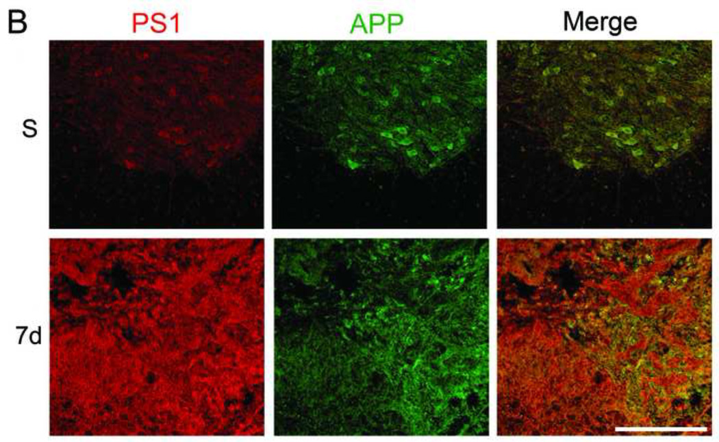
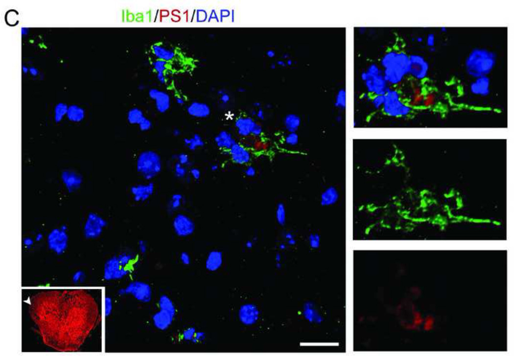
Figure 1. SCI causes an increase in PS1 and APP co-localization.
Mice were injured at T9 and sacrificed at 1, 3, and 7 days after contusion injury (n=4/group). The spinal cord sections were stained with APP (green) and PS1 (a component of γ-secretase, red) and confocal tile images were taken with a 20× objective. A. Representative images from sham (S, n=4) and 1, 3, and 7 days post-injury (DPI). PS1 and APP are both up-regulated after injury. B. The areas identified with arrowhead (in sham) and asterisk (injured, 7 days) in A are magnified to show the co-localization of APP and PS1 before and after injury, as evident by the increase in the overlap of red and green in the merged images at 7 days (Mag. Bar = 500 µm). C. Spinal cord sections from 1 DPI were stained with Ibal (green) and PS1 (red). The arrowhead in the 10× thumbnail image indicates the area from which the 63× confocal image was taken. The area identified by asterisk in the 63× was then digitally magnified (Mag. Bar =10 µm).


